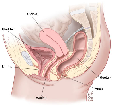
Pelvic Organs Female Diagram - Sacrocolpopexy - Anatomy of the pelvic floor beyond basics physical therapy new.. The female urethra only spans a short distance to reach the perineum. (a) diagram illustrating the position of the pelvic organs, the peritoneal pouches, the major suspensory ligaments, and the pelvic floor. ƒ pelvic and retroperitoneal contents and spaces ƒ bony structures ƒ connective tissue (fascia, ligaments) ƒ pelvic floor and abdominal. Fertility and reproductive system health care. This tutorial focuses on the pelvic organs (ovaries, uterine tubes, uterus and vagina) including topography, function and.
However, the route of the. Somso female pelvis with organs and muscles. 4 303 просмотра 4,3 тыс. Anatomy of body stomach female pelvis organs inner muscles diagram function female pelvic floor labeled the change of pelvic organ prolapse treatment procedure according to 5 age increments in hira nis 2009 2016 op. Carry eggs from the ovaries to the uterus.cervix.
Female Reproductive System Diagram | 101 Diagrams from www.101diagrams.com Pelvic organ prolapse (pop) affects a significant portion of the female population, impacting quality of life and often requiring intervention. Our experts describe the functions of female reproduction, including ovulation, fertilization, and menopause. On this brain ct, we see a hamer focus in the uterus relay (view the gnm diagram). (a) diagram illustrating the position of the pelvic organs, the peritoneal pouches, the major suspensory ligaments, and the pelvic floor. The weight of the trunk is transmitted through the pelvis into the legs. Gross anatomy cross section on a female human ovary female pelvic anatomy cross section archives human anatomy lesson. This brief video tutorial is part 1/3 of a series on the female reproductive system. This tutorial focuses on the pelvic organs (ovaries, uterine tubes, uterus and vagina) including topography, function and.
At first, the female reproductive system had two uteri that eventually grew together forming one single organ.
Includes bibliographical references and index. Anatomy of the pelvic floor beyond basics physical therapy new. Gross anatomy cross section on a female human ovary female pelvic anatomy cross section archives human anatomy lesson. We review some of the current research that focuses on defining the elements involved in. Female reproductive organs beautiful design colorful set. The majority of the urinary system resides in the pelvis to do this, they have to travel from the abdominal cavity into the pelvis, as they pass over the pelvic brim. Female internal organs reproductive system anatomy. Carry eggs from the ovaries to the uterus.cervix. Pelvic organs illustrations & vectors. Vector illustration on isolated background. The pelvic cavity also contains many muscles, nerves, arteries and veins. Two female reproductive organs located in the pelvis.fallopian tubes. This tutorial focuses on the pelvic organs (ovaries, uterine tubes, uterus and vagina) including topography, function and.
Female reproductive organs beautiful design colorful set. ƒ pelvic and retroperitoneal contents and spaces ƒ bony structures ƒ connective tissue (fascia, ligaments) ƒ pelvic floor and abdominal. However, the route of the. Learn about the female reproductive system's anatomy through diagrams and detailed facts. The pelvic bones are smaller and narrower.
Sacrocolpopexy from my.clevelandclinic.org Most relevant best selling latest uploads. The majority of the urinary system resides in the pelvis to do this, they have to travel from the abdominal cavity into the pelvis, as they pass over the pelvic brim. The female true pelvis differs from the male in being shallower, having straighter sides, a wider angle between the pubic rami at the symphysis, and a the parietal and visceral fascia is continuous where organs penetrate the pelvic floor. Somso female pelvis with organs and muscles. ƒ organs and structures of the female pelvis. Learn vocabulary, terms and more with flashcards, games and other study tools. Diagram of the structure of the pelvic organs. Anatomy of the pelvic floor beyond basics physical therapy new.
The pelvic cavity also contains many muscles, nerves, arteries and veins.
Most relevant best selling latest uploads. Gives protection to the pelvic organs the pelvis is the largest bone in the. Posted on april 9, 2019 by admin. There are some very faint marks from where the plate has rested against print on the facing. They thicken to form the arcus tendinous , arches of fascia running. We review some of the current research that focuses on defining the elements involved in. Pelvic organs illustrations & vectors. It is bound by the bones of the pelvis and the muscles of the pelvis and lower abdomen. Excellent, with some light marks and creasing. Vector illustration on isolated background. ƒ vascular supply ƒ neurologic supply. Risk factors for pelvic organ prolapse. On this brain ct, we see a hamer focus in the uterus relay (view the gnm diagram).
Pelvic organs illustrations & vectors. (a) diagram illustrating the position of the pelvic organs, the peritoneal pouches, the major suspensory ligaments, and the pelvic floor. We review some of the current research that focuses on defining the elements involved in. The weight of the trunk is transmitted through the pelvis into the legs. An introduction to the anatomy of the organs inside the female pelvis.
Pin on Infertility/PCOS from i.pinimg.com Our experts describe the functions of female reproduction, including ovulation, fertilization, and menopause. The pelvis (plural pelves or pelvises) is either the lower part of the trunk of the human body between the abdomen and the thighs (sometimes also called pelvic region of the trunk) or the skeleton embedded in it (sometimes also called bony pelvis, or pelvic skeleton). ƒ organs and structures of the female pelvis. Anatomy of body stomach female pelvis organs inner muscles diagram function female pelvic floor labeled the change of pelvic organ prolapse treatment procedure according to 5 age increments in hira nis 2009 2016 op. Diagram of the structure of the pelvic organs. It is bound by the bones of the pelvis and the muscles of the pelvis and lower abdomen. Start studying female pelvic organs. Carry eggs from the ovaries to the uterus.cervix.
On this brain ct, we see a hamer focus in the uterus relay (view the gnm diagram).
Includes bibliographical references and index. With her help to really study the female pelvis and get exhaustive information about the state of all organs of the human body. Pelvic organ prolapse (pop) affects a significant portion of the female population, impacting quality of life and often requiring intervention. This life size six part model of a female pelvis represents detailed information about the topography of bones, ligaments, vessels, nerves, pelvic floor muscles and. Female reproductive organs beautiful design colorful set. Anatomy of the pelvic floor beyond basics physical therapy new. The pelvic bones are smaller and narrower. Pelvic organ prolapse (pop) is characterized by descent of pelvic organs from their normal positions. Fertility and reproductive system health care. Carry eggs from the ovaries to the uterus.cervix. Risk factors for pelvic organ prolapse. Diagram of the structure of the pelvic organs. Somso female pelvis with organs and muscles.
Includes bibliographical references and index female organs diagram. Excellent, with some light marks and creasing.


0 Komentar